∷
∷
 ∷
∷
| NA Basic information |
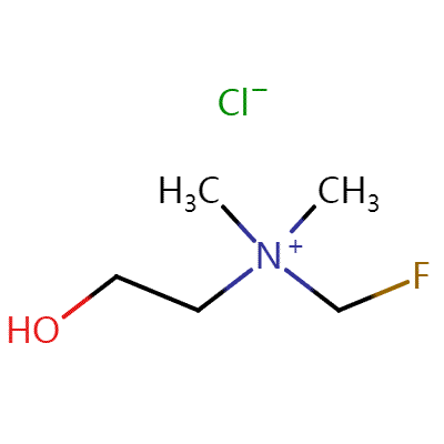
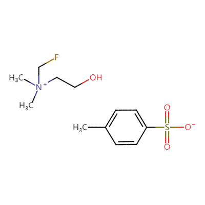
| Product Name: | OTS-choline fluoride |
| Synonyms: | |
| CAS: | 835605-47-5 |
| MF: | |
| MW: | C5H13FNO*C7H7O3S |
| EINECS: | |
| Product Categories: | |
| Mol File: | Mol File |
|
|
|
[European Radiology, 2023, vol. 33, # 1, p. 472 - 482]
[European Archives of Oto-Rhino-Laryngology, 2023, vol. 280, # 6, p. 2905 - 2910]
[Journal of Medical Imaging and Radiation Oncology, 2022, vol. 66, # 3, p. 324 - 331]
Value of 68Ga-labeled bombesin antagonist (RM2) in the detection of primary prostate cancer comparing with [18F]fluoromethylcholine PET-CT and multiparametric MRI—a phase I/II study
Abstract
Objectives: The bombesin derivative RM2 is a GRPr antagonist with strong binding affinity to prostate cancer (PCa). In this study, the impact of [68Ga]Ga-RM2 positron emission tomography-computed tomography (PET-CT) for the detection of primary PCa was compared with that of [18F]FCH PET-CT and multiparametric magnetic resonance imaging (mpMRI). Methods: This phase I/II study was conducted in 30 biopsy-positive PCa subjects. The patients were stratified into high (10 patients), intermediate (10 patients), and low risk (10 patients) for extraglandular metastases as defined by National Comprehensive Cancer Network (NCCN) criteria (NCCN Clinical Practice Guidelines in Oncology, 2016). The prostate gland was classified in 12 anatomic segments for data analysis of the imaging modalities as well as histopathologic findings. The segment with the highest radiotracer uptake was defined as the “index lesion.” All cases were scheduled to undergo prostatectomy with pelvic lymph node (LN) dissection in intermediate- and high-risk patients. Intraprostatic and pelvic nodal [68Ga]Ga-RM2 and [18F]FCH PET-CT findings were correlated with mpMRI and histopathologic results. Results: Of the 312 analyzed regions, 120 regions (4 to 8 lesions per patient) showed abnormal findings in the prostate gland. In a region-based analysis, overall sensitivity and specificity of [68Ga]Ga-RM2 PET-CT in the detection of primary tumor were 74% and 90%, respectively, while it was 60% and 80% for [18F]FCH PET-CT and 72% and 89% for mpMRI. Although the overall sensitivity of [68Ga]Ga-RM2 PET-CT was higher compared to that of [18F]FCH PET-CT and mpMRI, the statistical analysis showed only significant difference between [68Ga]Ga-RM2 PET-CT and [18F]FCH PET-CT in the intermediate-risk group (p = 0.01) and [68Ga]Ga-RM2 PET-CT and mpMRT in the high-risk group (p = 0.03). In the lesion-based analysis, there was no significant difference between SUVmax of [68Ga]Ga-RM2 and [18F]FCH PET-CT in the intraprostatic malignant lesions ([68Ga]Ga-RM2: mean SUVmax: 5.98 ± 4.13, median: 4.75; [18F]FCH: mean SUVmax: 6.08 ± 2.74, median: 5.5; p = 0.13). Conclusions: [68Ga]Ga-RM2 showed promising PET tracer for the detection of intraprostatic PCa in a cohort of patients with different risk stratifications. However, significant differences were only found between [68Ga]Ga-RM2 PET-CT and [18F]FCH PET-CT in the intermediate-risk group and [68Ga]Ga-RM2 PET-CT and mpMRT in the high-risk group. In addition, GRP-R-based imaging seems to play a complementary role to choline-based imaging for full characterization of PCa extent and biopsy guidance in low- and intermediate-metastatic-risk PCa patients and has the potential to discriminate them from those at higher risks. Key Points: • [68Ga]Ga-RM2 is a promising PET tracer with a high detection rate for intraprostatic PCa especially in intermediate-risk prostate cancer patients. • GRPr-based imaging seems to play a complementary role to choline-based or PSMA-based PET/CT imaging in selected low- and intermediate-risk PCa patients for better characterization and eventually biopsy guidance of prostate cancer disease.
The relationship between 18F-FCH uptake intensity and cell content in parathyroid lesions
Abstract
Purpose: To investigate the relationship between cell content and histopathological features of parathyroid lesions and 18F-FCH uptake intensity on PET/CT images. Methods: Patients with primary hyperparathyroidism (age > 18) who were referred to 18F-FCH PET/CT were involved. All patients underwent parathyroidectomy. Correlation of SUVmax with following factors were statistically analysed: serum PTH, Ca, P levels and histopathological parameters, total absolute amounts of chief cell, oxyphyllic cell and clear cell components calculated by the multiplication of the volume of the parathyroid lesion and the percentage of each type of cell content (called as Absolutechief, Absoluteoxyphyllic and Absoluteclear reflecting the total amount of each cell group). Results: A total of 34 samples from 34 patients (6M, 28F, mean age: 53.32 ± 15.15, min: 14, max: 84) who had a positive 18F-FCH PET/CT localizing at least one parathyroid lesion were involved. In the whole study group, SUVmax was found to be correlated with the greatest diameter and volume of the lesion and Absolutechief (p = 0.004, p = 0.002 and p = 0.035, respectively). In the subgroup analysis of 28 samples with longest diameter > 1 cm, the correlation between SUVmax and Absolutechief remained significant (p = 0.036) and correlation between SUVmax and volume and longest diameter became stronger (p = 0.011 and p > 0.001, respectively). No correlation was found between SUVmax and Absoluteoxyphyllic or Absoluteclear. Conclusions: There might be a relationship between 18F-FCH uptake intensity and chief cell content in patients with parathyroid adenoma. Further studies with larger patient groups would be beneficial to support the data.
Impact of pelvic dynamic acquisition on final reading of 18F-Fluorocholine positron emission tomography in patients with prostate adenocarcinoma: True need or unnecessary burden?
Abstract
Introduction: Despite the increasing use of 18F-fluorocholine (18F-FCH) positron emission tomography (PET) in patients with prostate cancer, the acquisition protocol remains debated. We have evaluated the influence of the pelvic dynamic phase on the final reading of whole-body 18F-FCH PET, to assess the need for a two-stage protocol. Reading the physician’s experience and patient’s previous treatment profile was also considered as potential influencing factors on final PET interpretation. Methods: All 18F-FCH PET/CT performed from January 2018 to September 2019 in patients with prostate cancer and including a pelvic dynamic phase followed by a delayed whole-body acquisition were retrospectively retrieved. PET/CT were analysed by one expert nuclear medicine physician and one resident. The whole-body scan was analysed blinded (first reading) and nonblinded from the results of the dynamic phase. Results: 221 consecutive PET/CT were selected from 201 patients previously treated by radical prostatectomy (n = 31), pelvic radiation therapy (n = 60), or both (n = 94). 24 patients had no previous treatments, and 12 benefited from other focal treatments. In the whole population, dynamic acquisition modified final interpretation of 32/221 scans (14.5%) for residents, 26 (11.8%) for experts and 19 (8.6%) for consensual reading. No influence of previous treatments was found. The availability of a dynamic phase would have been responsible for treatment modification in 5/221 scans (2.3%). Considering only the prostate bed, dynamic acquisition modified the final interpretation in 7/125 (5.6%) studies (consensual reading) from patients with previous prostatic surgery and 4/84 (4.8%) scans from patients without a history of prostatic surgical intervention. No significant influence of dynamic acquisition was found on the final PET interpretation on prostate lodge accordingly to previous prostatic surgery. Conclusion: The dynamic phase changes the interpretation of 18F-FCH PET in about 9% of cases and the therapeutic strategy in <3% of patients. The influence of the early phase reduces with physician experience. Patient's treatment profile does not appear to have a significant influence on the variability of interpretation, also including the prostate bed.
Contact: Mr Hsu
Phone: +86(0)1815701-8567
Tel: +86(0)570780-5618
Email: sales@fluorio.com
Add: No.5,Longfei Road,Longyou Eco-Tech Development Zone, Quzhou, Zhejiang, China